Mammographic Image Segmentation
Breast cancer is one of the leading cause of cancer deaths in women and the risk of developing breast cancer in life time for women is very high (8%-12%). Early detection is extremely important, since breast cancer can be successfully treated if diagnosed in the early stage. Mammography has been proved effective in examining abnormalities for early detection which is the key to improve breast cancer prognosis. Computer vision techniques have been widely used in digital mammogram analysis. Segmenting mass lesions is a critical step in automatic or computed aided detecting abnormalities and diagnosis.
Due to their natural handling of shape variation and independence of operation (once initialized), deformable contour models (snakes) are appropriate for the segmentation of mass lesions in digital mammographic images. The extracted shape and texture information through contour based segmentation are useful in determining benignancy or malignancy. In this work, we investigate four important edge-based active contour models for mass lesion segmentation. Two of them are classic active contour models and the other two are most recent advances in active contouring. Experiments are conducted on both synthetic and real images to compare their segmentation accuracy, and the ability in handling weak edges and difficult initializations.
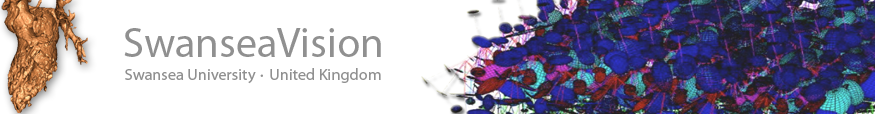
Example results - mass lesion segmentation
| Initial Snake | Evolving Snakes | Stabilized Snake | |
| Geodesic | 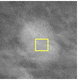 |  | 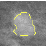 |
| GGVF |  | 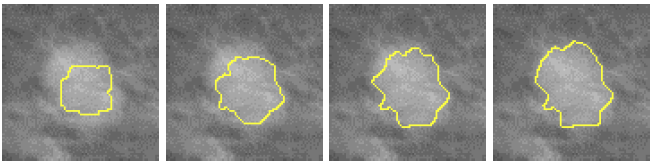 | 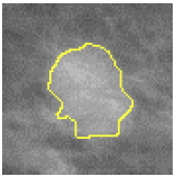 |
| GeoGGVF | 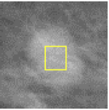 |  | 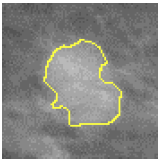 |
| MAC | 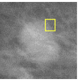 | 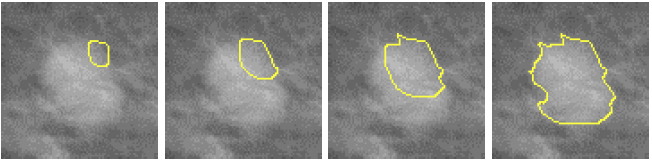 | 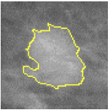 |
Example results - mass lesion segmentation (inconsistent edge contrast around the lesion & busier texture)
| Initial Snake | Evolving Snakes | Stabilized Snake | |
| Geodesic | 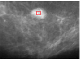 | 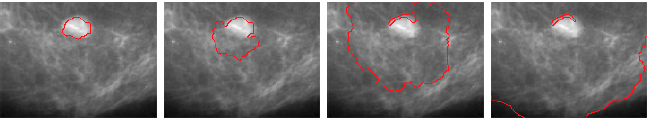 | 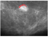 |
| GGVF | 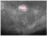 | 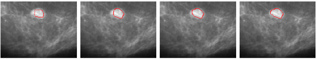 | 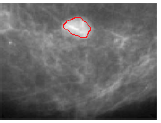 |
| GeoGGVF | 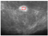 | 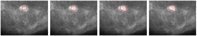 | 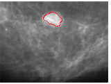 |
| MAC | 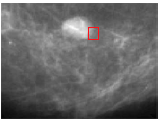 | 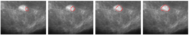 | 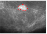 |
Example results - mass lesion segmentation - more results
| Original images | 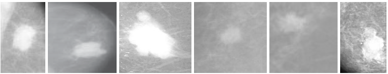 |
| Manual labeling | 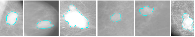 |
| Geodesic | 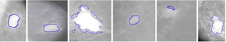 |
| GGVF | 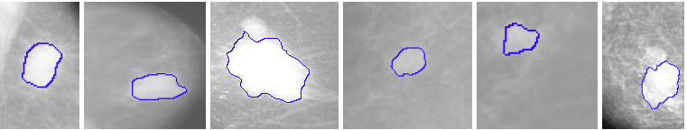 |
| GeoGGVF | 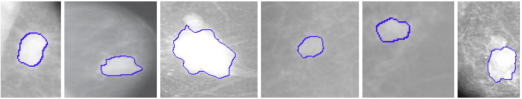 |
| MAC | 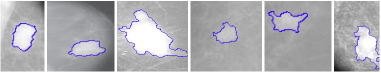 |
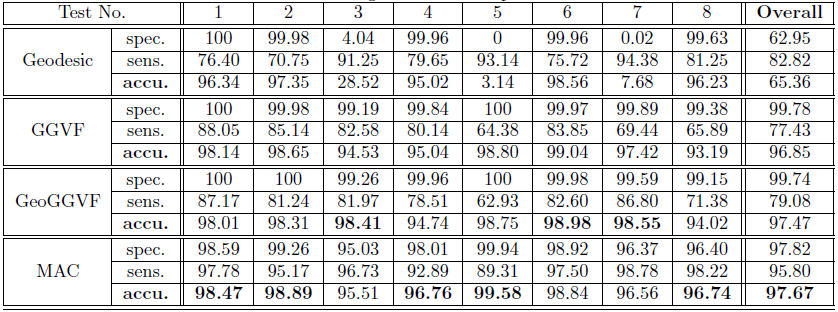
Example results - mass lesion segmentation - quantitative results
| The quantitative segmentation results. |
 |
Publications
- Xianghua Xie, An Applied Comparative Study on Active Contour Models in Mammographic Image Segmentation, In Performance Evaluation of Breast Cancer Screening, Diagnosis and Treatment, Edited by Ng, Rajendra Acharya, Suri, ASP, June, 2010.
- Xianghua Xie, Mammographic Image Segmentation using Edge Base Deformable Contour Models, In Proceedings of International Conference on Computational & Mathematical Biomedical Engineering, June 2009.