Intra-vascular Ultrasound (IVUS) imaging is a catheter-based technology, which shows 2D cross-sectional images of the coronary artery. In this work, we present a shape prior based graph cut method for IVUS image segmentation, which does not require user initialisation. The shape prior is generalised from multiple training shapes, rather than using singular templates as priors. Weighted directed graph construction is used to impose geometrical and smooth constraints learned from priors. The proposed cost function is built upon combining selective feature extractors. A SVM classi er is used to determine an optimal combination of features in presence of calcification, brotic tissues, soft plaques, and metallic stent, each of which has its own characteristics in ultrasound images. Comparative analysis on manually labelled ground-truth shows superior performance of the proposed method compared to conventional graph cut methods.
IVUS Image Structure
A typical IVUS image consists of lumen, vessel that includes intima and media layers, and adventitia that surrounds the vessel wall. The media-adventitia border represents the outer coronary arterial wall located between the media and adventitia. The media layer exhibits as a thin dark layer in ultrasound and has no distinctive feature. It is surrounded by brous connective tissues called adventitia. The appearance of the media-adventitia border in IVUS is a ected by various forms of artifact, such as acoustic shadow which can be caused by catheter guide wire, dense brous tissue or calci cation.
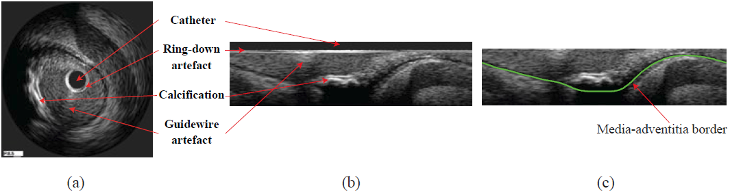 |
| An IVUS image (a) and its pre-processing: polar transformed image (b) and after removing the catheter region (c). |
Proposed Method
The images are rst transformed from Cartesian coordinates to polar coordinates and the catheter regions are removed (see above). This transformation not only facilitates the feature extraction and classi cation but also transfers a closed contour segmentation to a 'height-field' segmentation. The border to be extracted intersects once and once only with each column of pixels. This particular form of segmentation allows us to construct a node-weighted directed graph, on which a minimum path can be found without any user initialisation.
In non-prior graph construction the inter-column maximum distance is set as a constant. For our prior model, inter-column change should be in influenced by the derived shape prior. In calculating the shape prior cost function, the training shapes are aligned to our initial graph cut. The inter-column changes are then generalised using mean and standard deviation at individual column. These statistics are then used in determining maximum and minimum distances when connecting neighbouring columns in graph construction. Note that these inter-column arcs alone will impose a hard constraint on shape regularisation.
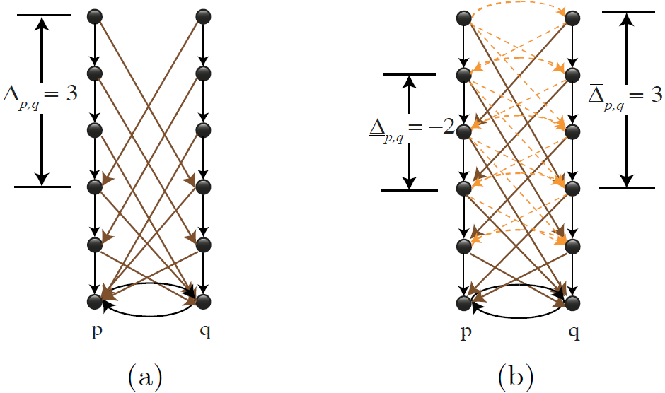 |
| Graph construction without shape prior where shape constraint is a global constant (a) and with shape prior model (b). |
Example results - comparison with s-t cut
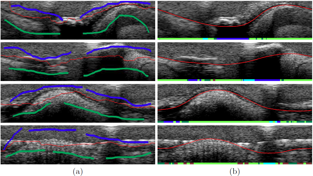 |
| Comparative results using (a) s-t cut (red) with user initialization(object: blue, background: green), and (b) proposed method. The bottom of each image also shows the classifi cation result: calci ed plaque (blue), brotic plaque (dark green), stent (dark red), guide-wire shadowing (cyan), and soft plaque/normal tissue (light green). |
Example results - comparison with groundtruth data
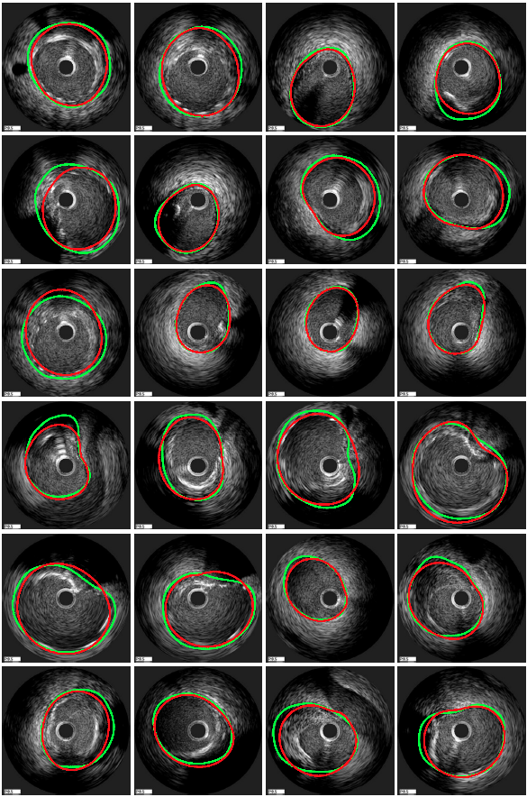 |
| Comparison between groundtruth (green) and the proposed method (red). |
Funding
This project was funded by the Welsh Government NISCHR under grant HA09/035.
Publications
- E. Essa, X. Xie, I. Sazonov, P. Nithiarasu, D. Smith, Shape Prior Model for Media-Adventitia Border Segmentation in IVUS using Graph Cut, In MICCAI Medical Computer Vision, September 2012.
- E. Essa, X. Xie, I. Sazonov, P. Nithiarasu, D. Smith, Graph-based Segmentation of Optimal IVUS Media-Adventitia Border using Shape Prior, In Proceedings of the 16th Conference on Medical Image Understanding and Analysis, July 2012.
- E. Essa, X. Xie, I. Sazonov, P. Nithiarasu, Automatic IVUS Media-Adventitia Border Extraction using Double-Interface Graph Cut Segmentation, In Proceedings of the 18th IEEE International Conference on Image Processing, September 2011.
- E. Essa, X. Xie, I. Sazonov, P. Nithiarasu, D. Smith, Automatic Segmentation of IVUS Media-Adventitia Border with Shape Prior, In Proceedings of the 15th Medical Image Understanding and Analysis, July 2011.